

Every child deserves the opportunity to stand, walk, and explore the world confidently. However, some babies are born with a foot deformity known as Congenital Talipes Equinovarus (CTEV) or Clubfoot, which can affect their ability to walk if left untreated. Fortunately, modern medical treatment and orthotic management have made CTEV one of the most successfully treatable congenital musculoskeletal conditions.
With early diagnosis, proper casting, appropriate orthotic intervention, and regular follow-up, most children with CTEV can achieve near-normal foot function and lead active, independent lives.
At Proactive Technical Orthopaedics, we believe that educating parents is just as important as providing the right orthotic care. This guide explains everything you need to know about CTEV, its causes, treatment options, and the role of orthoses in preventing recurrence.
What is CTEV (Clubfoot)?
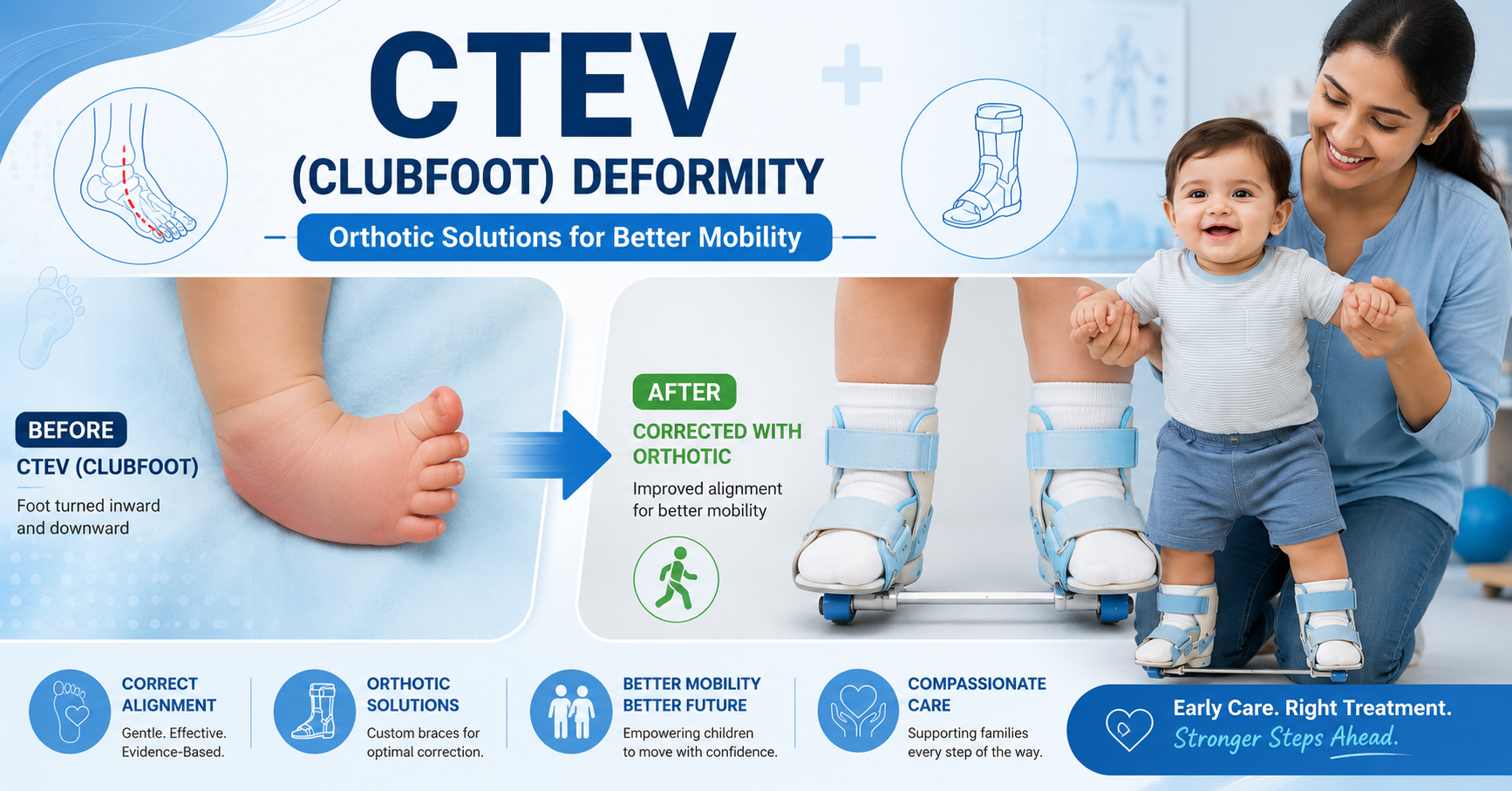
Congenital Talipes Equinovarus (CTEV), commonly known as Clubfoot, is a congenital deformity where one or both feet are twisted inward and downward. It develops before birth due to abnormal positioning of the bones, muscles, tendons, and ligaments of the foot and ankle.
The affected foot appears:
- Turned inward
- Pointed downward
- Smaller than a normal foot
- Stiffer than usual
- Difficult to move into a normal position
Clubfoot may affect:
- One foot (Unilateral Clubfoot)
- Both feet (Bilateral Clubfoot)
It occurs in approximately 1–2 babies per 1,000 live births, making it one of the most common congenital orthopaedics deformities worldwide.
Understanding the Four Components of CTEV
CTEV consists of four characteristic deformities, commonly remembered by the acronym CAVE:
C – Cavus
The arch of the foot becomes excessively high due to tight plantar structures.
A – Adduction
The forefoot deviates inward toward the midline of the body.
V – Varus
The heel tilts inward, making weight-bearing difficult.
E – Equines
The ankle remains in a downward-pointing position because of a tight Achilles tendon.
All four deformities must be corrected systematically for successful treatment.
What Causes Clubfoot?
The exact cause remains unknown in many cases. However, several factors are believed to contribute.
Genetic Factors
Children with a family history of clubfoot have a higher risk of developing the condition.
Neuromuscular Disorders
Clubfoot may occur with conditions such as:
- Spina bifida
- Arthrogryposis
- Cerebral palsy
Environmental Factors
Research suggests possible associations with:
- Smoking during pregnancy
- Reduced amniotic fluid
- Poor fetal positioning
In most cases, however, clubfoot occurs without any identifiable cause.
Signs and Symptoms
Parents often notice the deformity immediately after birth.
Common signs include:
- Foot turned inward and downward
- Small calf muscles
- Tight Achilles tendon
- High medial arch
- Shortened foot length
- Limited ankle movement
- Difficulty placing the foot flat on the ground
Without treatment, children may eventually walk on the outer border of the foot rather than the sole.
Diagnosis of CTEV
Diagnosis is usually straightforward.
Prenatal Diagnosis
Many cases can be detected during the 18–22-week ultrasound scan.
After Birth
Doctors perform:
- Physical examination
- Foot flexibility assessment
- Pirani Score
- Dimeglio Classification
- X-rays (only when necessary)
Early diagnosis allows treatment to begin within the first few weeks of life, when tissues are most flexible.
The Gold Standard Treatment: Ponseti Method
Today, the Ponseti Method is considered the worldwide gold standard for treating idiopathic clubfoot.
It involves four stages:
1. Gentle Manipulation
The foot is gradually stretched into a corrected position.
2. Serial Casting
Weekly plaster casts are applied for approximately 5–8 weeks to progressively correct the deformity.
3. Achilles Tenotomy
Most babies require a minor procedure to release the tight Achilles tendon, allowing proper ankle dorsiflexion.
4. Bracing (Orthotic Management)
After correction, an orthosis is essential to maintain the corrected position and prevent recurrence.
This final stage is often the most important because recurrence is common if bracing is not followed consistently.
Why Orthotic Management is Essential
Many parents mistakenly believe treatment is complete once casting ends. In reality, orthotic management plays a critical role in maintaining correction.
Without proper bracing:
- The foot may gradually return to its deformed position.
- Muscles and tendons can tighten again.
- Additional casting or surgery may become necessary.
Studies have shown that adherence to brace protocols significantly reduces the risk of relapse.
Orthotic Solutions for CTEV
Orthotic devices are prescribed according to the child’s age, stage of treatment, and clinical findings.
1. Foot Abduction Brace (FAB)
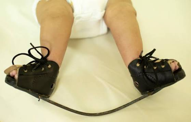
The Foot Abduction Brace is the most commonly used orthosis after Ponseti correction.
It consists of:
- Two open-toe shoes
- Adjustable connecting bar
- External foot rotation
- Proper dorsiflexion positioning
The brace keeps both feet in the corrected alignment while preventing relapse.
Wearing Schedule
Typically recommended as:
- 23 hours/day for the first 3 months after correction.
- Night-time and nap-time wear (12–14 hours/day) until 4–5 years of age, depending on the orthopedic surgeon’s advice.
2. Denis Browne Splint
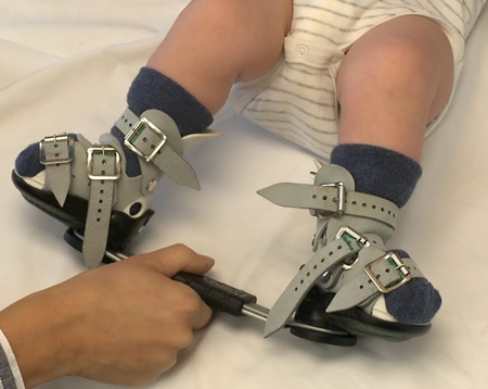
This traditional orthotic device connects both shoes with a rigid bar.
Its benefits include:
- Maintaining foot abduction
- Preventing inward rotation
- Supporting corrected alignment
Although newer brace designs are available, the Denis Browne splint remains effective when properly fitted.
3. Custom-Made AFO (Ankle Foot Orthosis)
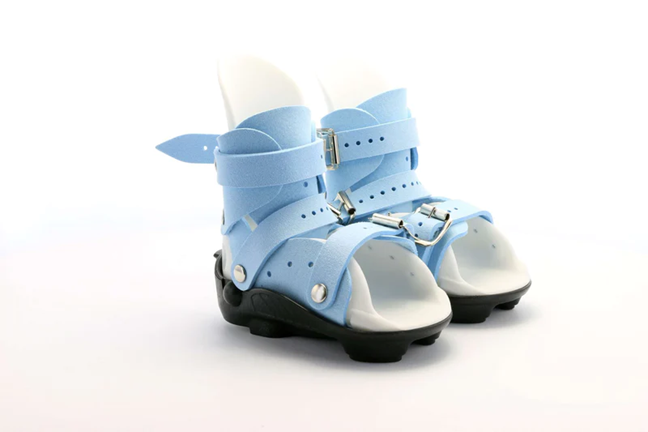
Some children—especially those with neurological conditions, recurrent deformity, or muscle imbalance—may benefit from a custom Ankle Foot Orthosis (AFO).
A custom AFO can:
- Improve ankle stability
- Support proper foot positioning
- Assist gait training
- Enhance balance and walking efficiency
Importance of Physiotherapy
Orthotic treatment works best when combined with physiotherapy and home exercise programs.
Physiotherapy focuses on:
- Stretching tight muscles
- Improving ankle mobility
- Strengthening lower-limb muscles
- Balance training
- Gait re-education
- Functional mobility exercises
Parents are also taught gentle stretching exercises to perform safely at home.
Conclusion
Congenital Talipes Equinovarus (CTEV) is a highly treatable condition when managed early with the right combination of the Ponseti Method, orthotic intervention, physiotherapy, and regular follow-up. Orthotic devices such as the Foot Abduction Brace, Denis Browne Splint, and custom AFOs are essential in maintaining correction and preventing relapse.
At Proactive Technical Orthopaedics, we believe that every child deserves the chance to walk, play, and grow without limitations. Through expert orthotic care, personalized rehabilitation, and family-cantered support, we help children with clubfoot take confident steps toward a healthier and more active future.